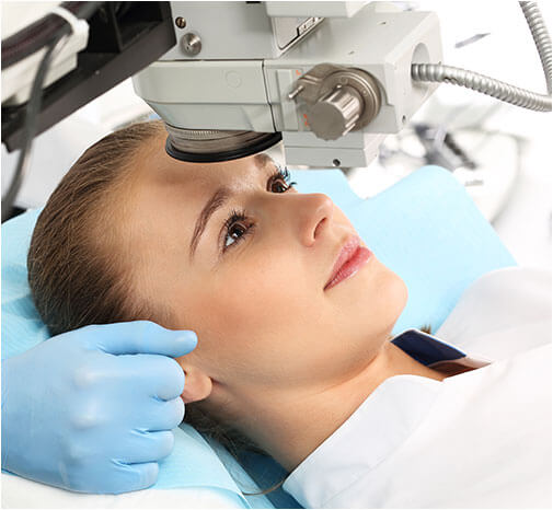
Retinal Photocoagulation

Retinal Photocoagulation
Overview
Background
Photocoagulation uses light to create a thermal burn in retinal tissue. When energy from a strong light source is absorbed by the retinal pigment epithelium (RPE) and is converted into thermal energy, coagulation necrosis occurs with denaturation of cellular proteins as temperature rises above 65°C.
Since the Diabetic Retinopathy Study (DRS), the source of light for photocoagulation has evolved from using a diffuse Xenon arc to using well-focused laser in the treatment of proliferative diabetic retinopathy. Presently, laser retinal photocoagulation is a therapeutic option in many retinal and eye conditions.
Effective retinal photocoagulation requires an unobscured view of the retinal tissue for light to be absorbed by pigment in the target tissue. In retinal tissue, light is absorbed by melanin, xanthophyll or haemoglobin. Melanin absorbs green, yellow, red and infrared wavelengths; xanthophyll (in the macula) absorbs blue but minimally absorbs yellow or red wavelengths; haemoglobin absorbs blue, green and yellow with minimal red wavelength absorption.
Indications
Indications for retinal photocoagulation include the following:
- Panretinal photocoagulation (PRP) for neovascular proliferative diseases such as proliferative diabetic retinopathy, sickle cell retinopathy, and venous occlusive diseases (see image below)
Panretinal photocoagulation in venous occlusive eye diseases. Image courtesy of National Eye Institute, National Institutes of Health.
- Focal or grid photocoagulation for macular oedema from diabetes or branch vein occlusion (see image below)
Focal or grid photocoagulation in macular oedema from diabetes or branch vein occlusion. Image courtesy of National Eye Institute, National Institutes of Health.
- Treatment of threshold and high-risk prethreshold retinopathy of prematurity
- Closure of retinal microvascular abnormalities such as microaneurysms, telangiectasia and perivascular leakage
- Focal ablation of extrafoveal choroidal neovascular membrane
- Creation of chorioretinal adhesions surrounding retinal breaks and detached areas [11]
- Focal treatment of pigment abnormalities such as leakage from central serous chorioretinopathy
- Treatment of ocular tumours
- Treatment of the ciliary body to decrease aqueous production for glaucoma
Preparation
Anaesthesia
Patients require anaesthesia for the procedure. Most patients undergo retinal laser procedures under topical anaesthesia such as proparacaine eye drops. Other patients will require subconjunctival, peribulbar, or retrobulbar injections of lidocaine.
Monitored aesthesia care or general anaesthesia is usually used for premature infants (with retinopathy of prematurity), children, and patients with problems in compliance.
Equipment
The laser source is connected via a fibreoptic cable to different types of delivery systems. Laser is delivered to the retina externally either through the cornea (transcorneal) or the sclera (transscleral). Transcorneal delivery employs a slit lamp (see first image below) or a Laser Indirect Ophthalmoscope (LIO). With the slit lamp delivery system, the laser is fired onto the retina using a contact lens which is placed on the corneal surface of the patient. Using the LIO delivery system, a noncontact binocular indirect ophthalmoscope condensing lens such as 28 D or 20 D lens (see second image below), is used to focus the laser onto the retina.
Slit lamp examination. Image courtesy of National Eye Institute, National Institutes of Health.
Laser indirect ophthalmoscopy (LIO). Image courtesy of National Eye Institute, National Institutes of Health.
Transscleral delivery uses a diode transscleral laser probe applied onto the sclera to treat the retina or ciliary body.
Laser can also be delivered internally (inside the eye), usually during vitrectomy procedures. An endolaser probe is introduced into the vitreous cavity, and laser is fired directly to the retina. The procedure is viewed using vitrectomy lens under an operating microscope.
Positioning
When using the slit lamp delivery system, the procedure is performed with the patient in a sitting position. With the endolaser and transscleral delivery systems, the patient is supine. With the LIO, the patient may be sitting or supine.
Complication Prevention
Proper laser protection goggles are required for all staff assisting the procedure. The laser safety filter (specific for each wavelength of laser) on the delivery system should always be activated upon performing the procedure.
The patient should be well positioned and instructed prior to the procedure. Retrobulbar block or general anaesthesia may be done for compliance problems.
The procedure should be performed or supervised by an experienced ophthalmologist to avoid technical errors resulting in complications from the procedure.
Technique
When using the slit lamp delivery system, a slit lamp contact lens is used to focus a beam of laser light onto the retina. With the indirect ophthalmoscope system, an indirect lens is used to focus the laser light onto the retina. With the endolaser, a laser probe is introduced into the vitreous cavity (usually during vitrectomy surgery) and the laser light is directly applied to the retina.
Modifications
Conventional laser delivery systems for retinal photocoagulation deliver spots individually on the retina. Newer semiautomatic laser delivery systems like the pattern scanning laser (PASCAL) have been designed to produce multiple spots on the retina in the same amount of time as conventional laser delivery systems. This makes the procedure less tedious and time consuming, allowing for better patient comfort.
New technology has been developed to minimize retinal damage, delivering micropulses of laser (micropulse laser). These micropulses have been shown to cause less retinal injury.
Post-Procedure
Complications
Although proven safe, like any other surgical procedure, retinal photocoagulation may occasionally be associated with complications. Before undergoing retinal photocoagulation, the patient should be fully informed of these, which include the following:
- Anterior segment complications such as corneal or lenticular opacification
- Transient visual loss
- Photocoagulation of the fovea
- Macular oedema
- Haemorrhage
- Choroidal Effusion
- Colour vision alterations
- Visual field defects and night vision problems
- Hemeralopia
