OPTICAL COHERENCE TOMOGRAPHY

OPTICAL COHERENCE TOMOGRAPHY (OCT)
Most people experience a change in their vision by the time they reach their early 40s. This can be a mild change in normal vision due to ageing or it could be a symptom of something more serious. An OCT scan can generate a highly detailed 3D image of the retina which will allow us to have a better view of your eye health.
Optical coherence tomography (OCT) is a noncontact, non-invasive imaging test. OCT uses light waves to take cross-section pictures of your retina.
With OCT, your ophthalmologist can see each of the retina’s distinctive layers. This allows your ophthalmologist to map and measure their thickness. These measurements help with diagnosis. They also provide treatment guidance for glaucoma and diseases of the retina. These retinal diseases include age-related macular degeneration (AMD) and diabetic eye disease.
The OCT is an excellent way to visualize the different layers of the retina and optic nerve in a living eye. Previous to these amazing technologies we were only able to see this view of an eye with tissue sections after the patient was deceased or if the eye was removed. The OCT allows us to diagnose and manage various eye problems like glaucoma and macular degeneration with greater accuracy and sensitivity
In OCT, cross-sectional images of the cornea and anterior segment are produced with optical backscattering of light in a fashion analogous to B-scan ultrasonography.
What happens during an OCT scan?
An OCT scan is very quick to perform and is completely painless and non-invasive. Results are available instantaneously and is a great way for patients to gain a better understanding of their eye condition. Just like a photo, the equipment will produce a 3D image of your retina in less than 15 seconds.
To prepare you for an OCT exam, your ophthalmologist may put dilating eye drops in your eyes. These drops widen your pupil and make it easier to examine the retina.
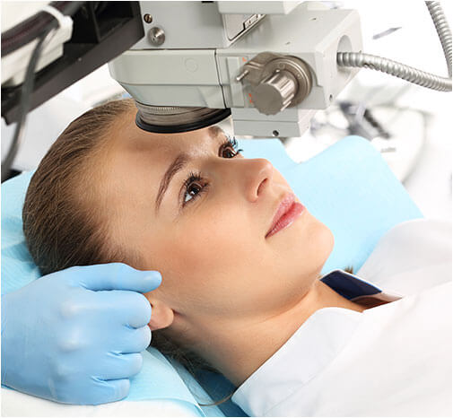
You will sit in front of the OCT machine and rest your head on a support to keep it motionless. The equipment will then scan your eye without touching it. Scanning takes about 5 – 10 minutes. If your eyes were dilated, they may be sensitive to light for several hours after the exam.
After examining the scans, your optometrist will discuss the results with you. We store the scans in your records to compare the images with future images of your retina.
What conditions can an OCT scan help diagnose?
There are many conditions of the eye which can be diagnosed with the help of an OCT scan. Serious eye disease can develop in the retina as we age and these are often not visible on the surface of the eye or exhibit physical symptoms. Early detection is our best ally in treating these diseases and an OCT scan is the best way to monitor the health of your retina.
OCT is useful in diagnosing many eye conditions, including:
- macular hole
- macular pucker
- macular edema
- age-related macular degeneration
- glaucoma
- central serous retinopathy
- diabetic retinopathy
- vitreous traction
OCT is often used to evaluate disorders of the optic nerve as well. The OCT exam helps your ophthalmologist see changes to the fibres of the optic nerve. For example, it can detect changes caused by glaucoma.
OCT relies on light waves. It cannot be used with conditions that interfere with light passing through the eye. These conditions include dense cataracts or significant bleeding in the vitreous.
